
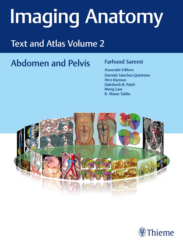
Imaging Anatomy: Text and Atlas Volume 2: Abdomen and Pelvis (Atlas of Imaging Anatomy)
Product details
| Management number | 231930532 | Release Date | 2026/06/18 | List Price | $64.86 | Model Number | 231930532 | ||
|---|---|---|---|---|---|---|---|---|---|
| Category | |||||||||
Unique anatomic atlas provides an indispensable virtual desk dissection experienceNormal imaging anatomy and variants, including diagnostic and surgical anatomy, are the cornerstones of radiologic knowledge. Imaging Anatomy: Text and Atlas Volume 2, Abdomen and Pelvis is the second in a series of four richly illustrated radiologic references edited by distinguished radiologist Farhood Saremi. The atlas is coedited by esteemed colleagues Damián Sánchez-Quintana, Hiro Kiyosue, Dakshesh B. Patel, Meng Law, and R. Shane Tubbs with contributions from an impressive cadre of international authors. Succinctly written text and superb images provide readers with a virtual, user-friendly dissection experience.This exquisitely crafted atlas combines fundamental core anatomy principles with modern imaging and postprocessing methods to increase understanding of intricate anatomical features. Twenty-two concise chapters cover the abdominal wall, alimentary tract, liver, biliary system, pancreas, spleen, peritoneum, genitourinary system, pelvic floor, neurovasculature, and surface anatomy. Relevant anatomical components of the abdomen and pelvis are discussed, including musculature, arteries, veins, lymphatics, ducts, and innervation.Key HighlightsHigh-quality cross-sectional multiplanar and volumetric color-coded CT, MRI, and angiography imaging techniques provide detailed insights on specific anatomyCross-sectional and topographic cadaveric views by internationally known anatomists coupled with more than 1,600 illustrations clearly elucidate difficult anatomical conceptsConsistently formatted chapters include an introduction, embryology, review of anatomy, discussion of anatomical variants, postsurgical anatomy, and congenital and acquired pathologiesThis unique resource provides an excellent desk reference for differentiating normal versus pathologic anatomy. It is essential reading for medical students, radiology residents and veteran radiologists, internists, and general surgeons, as well as vascular and transplant surgeons. Read more
| ASIN | B0BHZWPQLX |
|---|---|
| XRay | Not Enabled |
| ISBN13 | 978-1638536116 |
| Edition | 1st |
| Language | English |
| File size | 351.7 MB |
| Page Flip | Enabled |
| Publisher | Thieme |
| Word Wise | Not Enabled |
| Book 2 of 3 | Atlas of Imaging Anatomy |
| Print length | 664 pages |
| Accessibility | Learn more |
| Screen Reader | Supported |
| Publication date | October 14, 2022 |
| Enhanced typesetting | Enabled |
Bestseller ranking
Digestive Organs





Correction of product information
If you notice any omissions or errors in the product information on this page, please use the correction request form below.
Correction Request Form